Histology
Lymph node biopsy
A biopsy is the withdrawal of tissue from a living organism. In order to confirm the presence of a malignant lymphoma, it is indispensable to perform a biopsy of a complete lymph node.
The lymph node chosen for this should be easily accessible and as large as possible. In most cases, the biopsy can be performed under local anesthesia, but if the mediastinum or the abdomen are involved, a general anesthesia is required.
After its removal, a histological examination will be performed on this lymph node, including cell staining and a microscopic analysis. Through this process the pathologist can identify characteristic signs of the disease such as the typical Hodgkin cells and Reed-Sternberg cells in case of classical Hodgkin lymphoma.
Whenever possible, the diagnosis should be reviewed and confirmed by a second pathologist who is particularly experienced in lymphoma diagnostics, a review pathologist. Within the trials of the GHSG this is a standard procedure.
Histology of tumor cells
The neoplastic Hodgkin lymphoma cells (the Hodgkin and Reed-Sternberg cells and their variants) have a typical morphology. The Hodgkin lymphoma infiltrate contains only few of these neoplastic cells (approx. 1%) but a large number of non-malignant cells, such as T- and Blymphocytes, macrophages and eosinophil granulocytes.
Hodgkin lymphoma can be diagnosed and classified by means of a histopathological tissue examination. The World Health Organization (WHO) distinguishes between classical Hodgkin lymphoma (approx. 95% of cases) and nodular lymphocyte-predominant Hodgkin lymphoma (approx. 5% of cases ). The latter is classified as a separate disease.
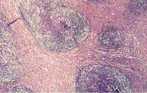
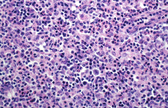
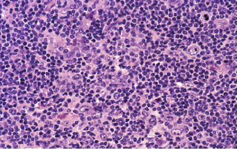
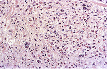
Classical Hodgkin lymphoma is divided into four histologic subtypes.
|
Histologic subtype |
% of cases |
image figure |
|
Nodular-sclerosing type |
65% |
|
|
Mixed cellularity type |
25% |
|
|
Lymphocyte-rich classical Hodgkin lymphoma |
4% |
|
|
Lymphocyte-depleted classical Hodgkin lymphoma |
1% |
|
The histologic subtypes of Hodgkin lymphoma do not influence the choice of treatment anymore today. However, stage IA nodular lymphocyte-predominant Hodgkin lymphoma, in which only one lymph node region is affected, has a very favorable course compared to the other types at stage I, and therefore treatment consists in involved-field radiotherapy only.


