Pathology review

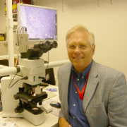
Prof. Dr. med. Dr. h.c. Harald Stein
Pathology review coordinator of the GHSG
Contact
Berliner Referenz- und Konsultationszentrum für Lymphom- und Hämatopathologie am Institut für Pathodiagnostik Berlin Komturstrasse 58-62 12099 BerlinDevelopment and organization
Reproducibility of histopathological diagnoses used to be rather unsatisfactory for decades. This was reflected in many different systems of classification that were inconsistent and incompatible. Due to new, groundbreaking insights into immunology in the 1970s and the following years, the situation improved. The following findings were essential for lymphoma classification and diagnostics:
- Lymphocytes are no terminally differentiated cells, but able to transform into blasts capable of reproduction as a result of certain signals.
- The lymphatic system mainly consists of two cell types, B cells and T cells, that have different origins and functions.
- B cells and T cells have numerous different forms with different functions.
Further decisive progress was made with the discovery of some morphological, immunophenotypical and genetic characteristics, with the help of which many different lymphatic diseases could be defined precisely based on their cellular origin. However, it became evident that in lymphatic malignancies a high level of diagnostic reproducibility could only be achieved if the mentioned complex diagnostic criteria were applied by pathologists specialized in lymphatic and hematologic pathology and primarily concerned with research and diagnostics of the lymphatic system’s diseases.
In order to achieve the highest possible level of correctness and diagnostic reproducibility for patients of the German Hodgkin Study Group, a pathology review panel consisting of four review pathologists was established at an early stage for reassessment of diagnostic results. During the first years (from 1984 to 1996) these reviews were carried out retrospectively in meetings of all four review pathologists who examined samples under the microscope together. The rapidly increasing number of recruited trial patients finally rendered this kind of pathology reviews impossible as they took too much time and effort. Therefore, the pathology review method was changed in 1996 to a prospective approach. Reassessments were performed promptly from then on (if possible, before start of treatment) and by one review pathologist only. At the same time, the number of review pathologists was raised to six. To ensure correctness and reproducibility of diagnostics among the review pathologists, all six review pathologists meet at regular intervals for joint microscopic examinations. During these meetings they mainly discuss cases in which primary pathologist and review pathologist have made different diagnoses. At first, each pathologist performs a separate microscopic examination. After that, a microscope that can be used by several people at the same time is employed for a joint evaluation. This procedure has proven to be very reliable and produces a high diagnostic consensus between the six review pathologists.
During the last 15 years, approximately 10,000 samples have been assessed by the review pathologists within the scope of the GHSG’s trials. In most cases their diagnosis is consistent with that of the primary pathologist. In 3-5% of all cases, however, a different type of lymphoma is diagnosed (e.g. NHL) or the presence of a malignant disease cannot be verified. These facts illustrate the importance of pathology reviews.
Diagnostic criteria
Pathology review diagnostics and Hodgkin lymphoma classification are based on the WHO lymphoma classification system of 2008. This implies that, in order to complement and confirm morphological diagnostics, immunohistological diagnostics have to be performed in addition. Immunohistological diagnostics include the following marker molecules:
Classical Hodgkin lymphoma: CD30+, CD15, LMP-1, PAX5, CD20 and CD3. In problematic cases, the following immune reactions are tested in addition: IRF4/MUM1, Oct2A and BOB.1, ALK, Perforin
Lymphocyte-predominant Hodgkin lymphoma CD20, Oct2A, J chain, CD30
Review pathologists:
- Prof. Dr. med. H. Stein, Berlin (Coordinator)
- Prof. Dr. med. W. Klapper, Kiel
- Prof. Dr. med. A. Rosenwald, Würzburg
- Prof. Dr. med. A. C. Feller, Lübeck
- Prof. Dr. med. M.-L. Hansmann, Frankfurt
- Prof. Dr. med. P. Möller, Ulm
- Prof. Dr. med. G. Ott, Stuttgart
- Prof. Dr. med. F. Fend, Tübingen